Principle of multiphoton excitation microscopy
General principle of two-photon excitation process
Two-photon (or multiple-photon) excitation microscopy is based on the principle that two (or more) infrared photons can excite a molecule the same way a single photon of double (or more) energy.
Confinement of excitation is achieved because of the high amount of photon generated by the infrared pulsed laser is focalized on the sample. The photon density is so high at this point that the probability that two photons interact with a molecule at the same location and at the same moment is different from zero.
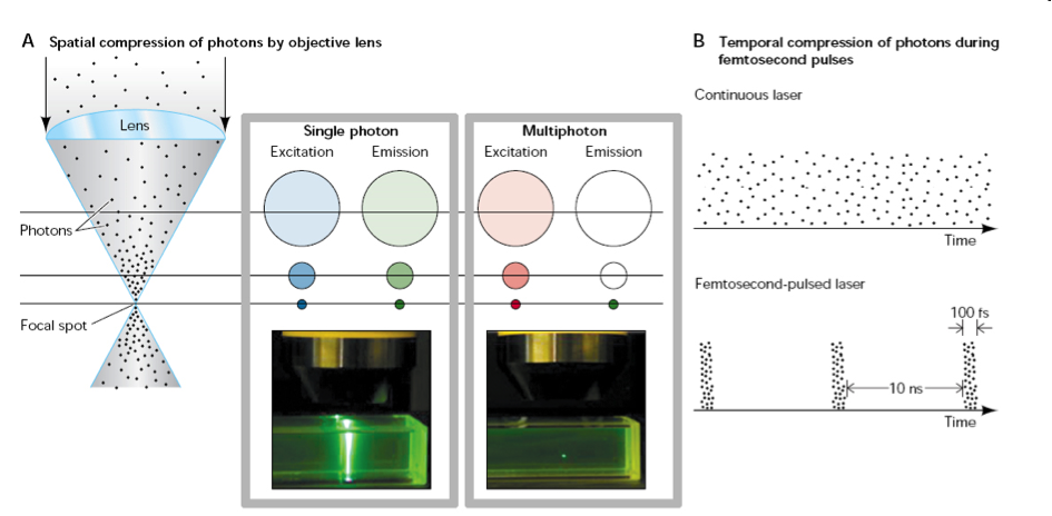
From The Parker Lab
Main advantages: localization of excitation implies no out-of-focus bleaching of fluorophores, no out-of-focus photodamaging, and no out-of-focus emitted fluorescence. This means also that all the emitted photons come from the focal point.
Infrared light diffuses well in the tissues so that microscopy enable achieving imaging of thicker samples than conventional visible light-excitation microscopy.
| Single photon excitation | Two-photon excitation |
|---|---|
| rlat = 0.51 λ / NA | rlat = 0.377 λ / NA (if NA ≤ 0.7) |
| rax = (0.88 λ ) / (n-√(n2-NA2)) | rlat = 0.383 λ / NA0.91 (if NA > 0.7) |
| rax = (0.626 λ ) / (n-√(n2-NA2)) |
λ = largest wavelength; n = refraction index of objective immersing medium; rlat = lateral resolution; rax = axial resolution. From James Pawley, 1995 and Warren Zipfel, 2003 (modified by Emmanuel Schaub)
Choice of the excitation wavelength
Opportunity window

Endogenous molecules absorb light. It is important to avoid wavelength that could excite water (blue line), hemoglobin (red and orange lines), melanin (black line), fat (green line) or tryptophan (pink line).
Definition of the two-photon excitation cross-section
The two-photon cross-section (σ2p) is a quantitative measure of the probability of a two-photon absorption. σ2p has units of cm4.s, with 10-50cm4 called a Göppert-Mayer or GM. Because it is difficult to measure σ2p directly, the two-photon action cross-section (δ2p) is usually measured; this is the product of the fluorescence quantum yield (ΦF) and the absolute two-photon absorption cross-section (σ2p). Both the wavelength dependence and the absolute values of ΦFσ2p are important in multiphoton microscopy. Warren Zipfel, et al., 2003
1 GM = 10-50cm4.s.photon-1
δ2p = Φσ2p
Examples:
- NAD(P)H, retinol, folic acid: δ2P < 10-4GM
- EGFP: 41.21GM at 920nm
- Qdots ≈ 50,000GM
- Some caged compounds between 10-2GM and 102GM
Cross-section of common fluorescent proteins
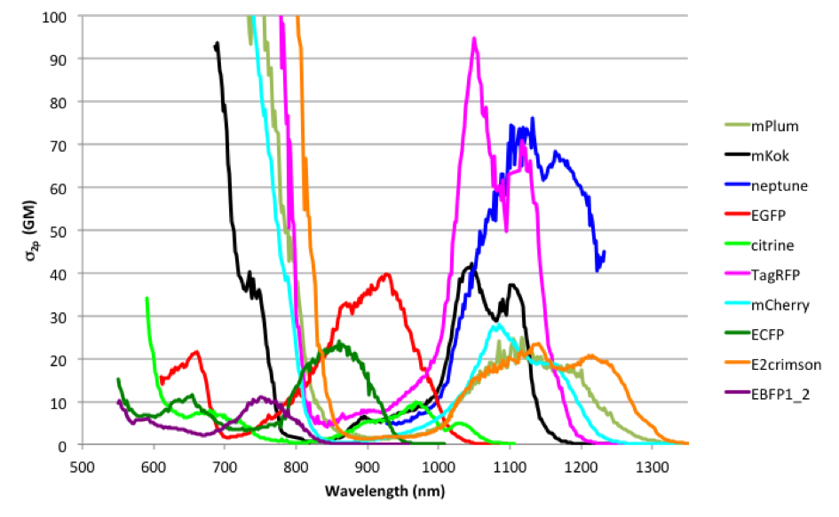
Compiled absorption spectra adapted from Mikhail Drobizhev, et al., 2011
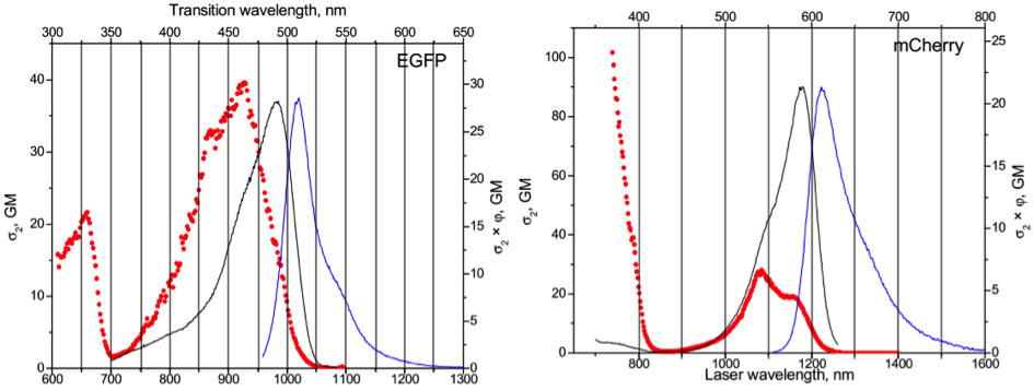
Absorption spectra of EGFP and mCherry adapted from Mikhail Drobizhev, et al., 2011
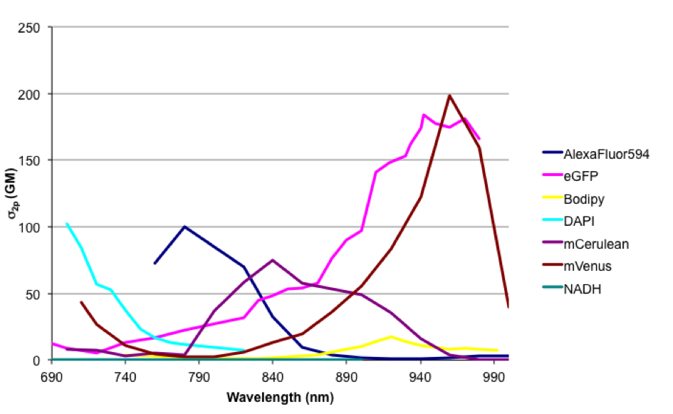
Cross-section of some fluorescent dyes
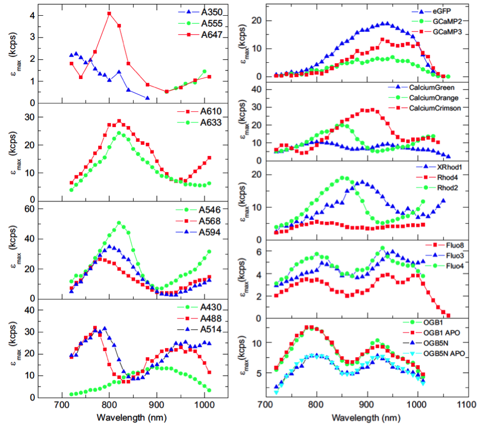
Compilation of absorption spectra of fluorescent dyes adapted from Jörg Mütze, et al., 2012
Cross-section from some other molecules
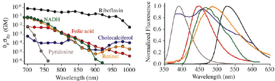
Absorption spectra of other molecules adapted from Warren Zipfel, et al., 2003
Compilation of absorption spectra from DRBIO